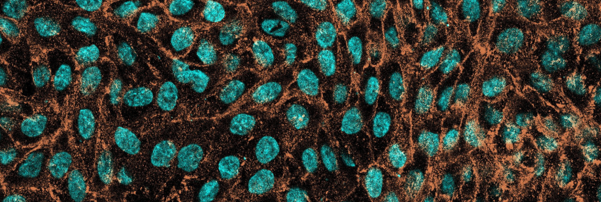
The Bioimaging Platform at IRTA-CReSA is a core facility fully integrated within a high-containment environment (NSB3). This unique setting places IRTA-CReSA among the few institutions worldwide offering advanced bioimaging services directly within a facility designed for work with high-risk pathogens, enabling research questions that would be unfeasible in conventional environments.
The High-Containment Unit at IRTA-CReSA is part of the RLABS node, a Spanish ICTS (Unique Scientific and Technical Infrastructure) for High-Containment Research. ICTSs are strategic, one-of-a-kind facilities providing high-level scientific services, ensuring maximum standards of safety, quality, and accessibility for the research community.
Thanks to this integration within an ICTS, the Bioimaging Platform at IRTA-CReSA is unique in Spain. It offers advanced imaging technology within an NSB3 environment, making it possible to carry out complex studies involving infectious agents with complete safety and regulatory compliance.
The members of the Bioimaging Platform provide assistance in experimental design, data acquisition, and analysis, as well as comprehensive training and technical support to ensure optimal use of our facilities and equipment. In short, the team offers a fully integrated service covering all phases of bioimaging research, providing continuous guidance to guarantee robust and reproducible results.
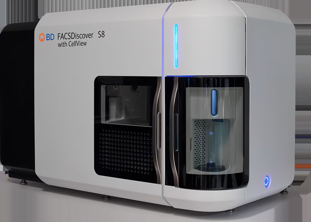
Our advanced equipment enables work with both live and fixed or cryopreserved samples, always maintaining optimal safety and integrity within the NSB3 facilities.
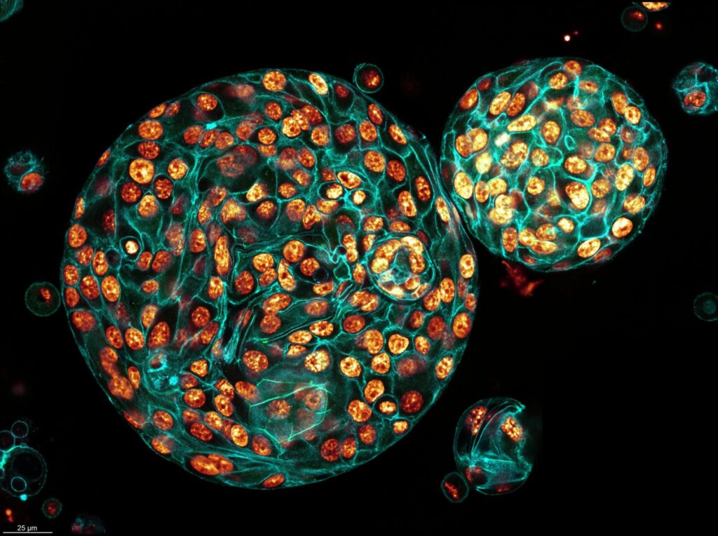
The Stellaris 8 confocal microscope is an inverted system optimized for live-cell imaging, equipped with an incubation chamber that maintains physiological conditions during extended acquisition sessions. It features a white light laser covering 440–790 nm and three HyD S photon-counting detectors, providing excellent sensitivity and enabling quantification of fluorescence lifetimes via the TauSense module.
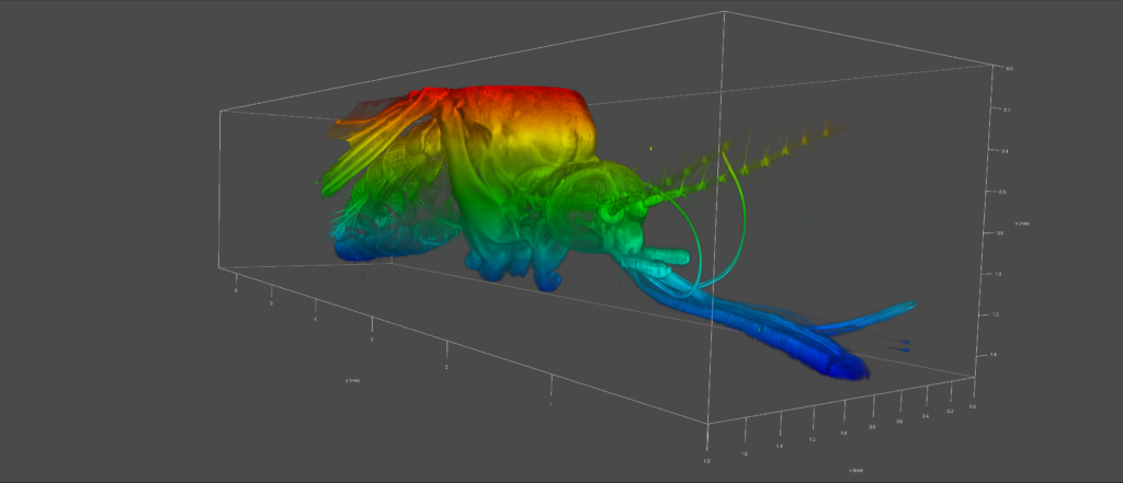
This system also includes the Digital LightSheet (DLS) module, which significantly expands its capabilities, allowing imaging of larger samples, both live and fixed/cleared, with improved penetration depth. The DLS module enables high-speed acquisition while minimizing photodamage, making it ideal for dynamic experiments and the generation of 3D maps with better sample preservation.
The IncuCyte SX5 is an automated imaging system integrated within an incubator, allowing continuous, non-invasive monitoring of cellular processes over time. This setup enables the study of biological dynamics under strictly controlled conditions without disrupting culture stability.
It is equipped with green, orange, and NIR fluorescence modules and includes advanced analysis tools, including AI-based segmentation modules. These tools allow quantification of proliferation, cell movement, morphology, interactions, and responses to infection with high precision and reproducibility.
At IRTA-CReSA, the system is fully integrated within the NSB3 environment, enabling real-time visualization of infected samples or samples containing high-risk pathogens. This capability makes dynamic infection studies possible under conditions that would be impossible in conventional settings, always ensuring safety and regulatory compliance.
To process samples without removing them from the NSB3 environment, the Bioimaging Platform has a Leica CM3050 cryostat that sections OCT-embedded samples while preserving tissue antigenicity. This is particularly valuable for infectious samples that cannot leave the containment area, providing high-quality sections for immunofluorescence and other applications.
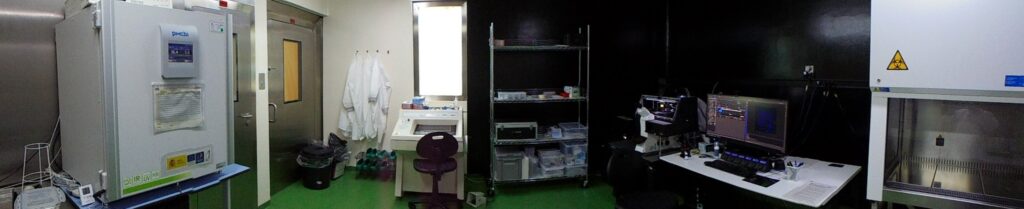
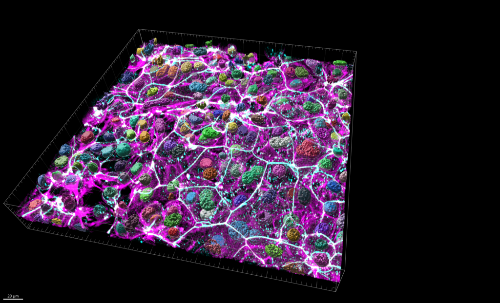
The platform includes a high-performance workstation designed to efficiently analyze the large volumes of data generated by the confocal, Digital LightSheet, and IncuCyte systems. This workstation features an Intel Xeon W7-2495X processor with 24 cores and 48 threads, an NVIDIA RTX A6000 GPU with 48 GB of GDDR6 memory, and 256 GB of DDR5 ECC RAM, providing the necessary power for complex 3D reconstructions, advanced quantification, and computationally intensive AI-based segmentation processes.
The workstation is equipped with Imaris 10.1, specialized software for 3D and 4D data visualization and analysis, as well as a broad set of open-source tools including Fiji/ImageJ, QuPath, Matlab, and other solutions for quantification, morphological analysis, and multichannel segmentation. Together, this creates a comprehensive and optimized environment to support high-complexity imaging projects.
Additionally, the platform has a centralized data storage server (NAS) of over 60 TB, connected via high-speed cabling to the imaging systems to ensure stable and efficient data flow. This server is accessible locally from the image analysis workstation and remotely through a secure web link from anywhere in the world, allowing researchers to view, download, or share data conveniently and flexibly, even outside the IRTA-CReSA facilities.
Joan Repullés: bioimaging@irta.cat